For decades, neuroscience suffered from a frustrating limitation. Researchers could record electrical activity from individual neurons or blast entire regions of tissue with electricity, but they lacked the ability to target specific cells within a living circuit with millisecond precision. It was akin to trying to understand how a complex computer works by dropping an anvil on the motherboard. Everything changed when a quiet revolution called optogenetics emerged, joining microbiology with fiber optics to manipulate the firing of neural circuits using pulses of light.
The architects of this transformation—Peter Hegemann, Georg Nagel, and Karl Deisseroth—were awarded the Nobel Prize in Physiology or Medicine for discoveries concerning light-gated ion channels. While mainstream headlines praised the honor as a victory for brain mapping, the reality inside active laboratories is far more complex. This recognition is not merely a celebration of a neat scientific trick; it marks the official transition of neuroscience from an observational science into a rigorous, causal discipline.
The Algal Origins of a Neural Switch
To understand why this breakthrough deserved a Nobel Prize, you have to look away from mammalian brains and down into a drop of pond water. Long before neuroscientists dreamed of controlling thought patterns with fiber optics, single-celled green algae like Chlamydomonas faced a fundamental evolutionary problem: they needed to swim toward sunlight to survive, but they had no eyes.
Peter Hegemann and Georg Nagel spent years investigating how these microscopic organisms sensed light. They discovered channelrhodopsins, specialized proteins embedded in the algal cell membrane that act as microscopic, light-sensitive gates. When struck by a specific wavelength of light, these channels instantly pop open, allowing charged ions to flood across the membrane and generate an electrical impulse within half a millisecond.
For years, plant physiologists studied these proteins as botanical curiosities. The monumental leap occurred when Karl Deisseroth, a psychiatrist and bioengineer at Stanford University, recognized that these microbial proteins could be transplanted into animal cells. If a mammalian neuron could be forced to manufacture a channelrhodopsin gene, that neuron would suddenly become sensitive to light. Shine a blue laser on the cell, and it fires an action potential. Turn off the light, and it goes quiet.
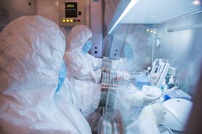
In a hypothetical laboratory setting, a researcher can inject a viral vector carrying the channelrhodopsin gene into a specific cluster of motor neurons in a mouse. By threading a microscopic hair-thin optical fiber into the skull, the investigator can deliver brief flashes of light, commanding the animal to move its whiskers or run in circles on command. The precision was absolute. Instead of exciting an entire brain structure indiscriminately, scientists could now isolate and manipulate a handful of cells out of millions.
Moving Past the Limits of Electrical Stimulation
Traditional electrophysiology relied on metal microelectrodes. While electrodes revolutionized our early understanding of action potentials, they suffer from a severe flaw: electrical current does not discriminate. When an electrode discharges inside brain tissue, it fires every neuron, axon, and passing fiber in its immediate vicinity. It is a blunt instrument.
Optogenetics solved this specificity crisis by turning genetic engineering into a remote-control switch. By attaching the light-sensitive protein to specific promoter genes, researchers could target only dopamine-producing neurons, or exclusively inhibitory interneurons, leaving neighboring cells completely unaffected.
Consider the study of psychiatric disorders like major depression or Parkinson's disease. For generations, clinicians knew that certain brain regions were broadly correlated with mood or movement, but proving a causal link remained elusive. Did an overactive circuit cause anxiety, or was it merely reacting to it? Optogenetics answered this by allowing scientists to switch specific circuits on and off in real time while observing behavioral changes in animal models. The technology shifted the entire field from correlation to direct causation.
The Clinical Bottleneck
Despite the euphoria surrounding the Nobel announcement, translational medicine faces a massive wall. Translating a technique that relies on genetic modification and implanted fiber-optic cables into human patients is an extraordinarily steep climb.
Viruses must be engineered to deliver the necessary genes safely into human tissue without triggering adverse immune responses. Furthermore, permanent fiber-optic implants inside the human skull carry risks of chronic inflammation, tissue scarring, and mechanical failure over time.
Progress is happening, albeit slowly. Clinical trials are currently exploring optogenetic therapies to restore partial vision in patients suffering from degenerative retinal diseases like retinitis pigmentosa, where photoreceptor cells have died, but downstream retinal neurons remain intact. By introducing light-gated channels into surviving inner retinal cells, researchers hope to bypass the broken phototransduction machinery entirely and project visual patterns directly onto the inner lining of the eye.
Yet, applying this technology to complex psychiatric conditions in humans—such as severe treatment-resistant depression or schizophrenia—remains far on the horizon. The human brain is exponentially larger and more intricate than a rodent model, and altering its genetic code permanently to insert algal proteins introduces regulatory and safety hurdles that will take decades to clear.
The Paradigm Shift in Neurotechnology
The 2026 Nobel Prize acknowledges that mapping the structural connectome of the brain—tracing every single synapse like a static road map—is fundamentally insufficient. A road map tells you where the roads go, but it does not tell you where the traffic is flowing or which intersections are jammed.
By giving researchers the ability to read and write neural activity simultaneously with light, Hegemann, Nagel, and Deisseroth provided the tools required to watch the living mind in action. The ultimate legacy of their work will not be measured by the prestige of Stockholm medals, but by how effectively future generations of scientists translate these optical switches into genuine cures for neurological disorders that have baffled medicine for centuries.
The path forward requires navigating immense biological hurdles, but the era of guessing how neural circuits operate has officially ended, replaced by an era where science can finally turn the lights on inside the mind.
Stanford professor wins Nobel Prize for discovery of new way to study brain using light
This video provides an overview of the research conducted by Karl Deisseroth at Stanford University that contributed to the 2026 Nobel Prize in Physiology or Medicine.