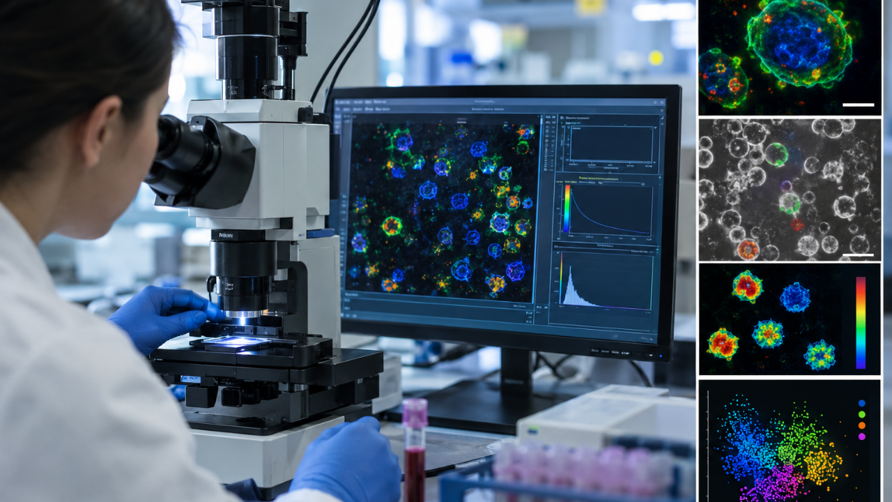
For decades, observing the immune system in action meant poisoning it first. Standard laboratory protocols rely heavily on fluorescent antibodies, chemical dyes, and exogenous markers to tag specific surface proteins, a process that alters cellular physiology and destroys cell viability. A recent optical breakthrough changes that dynamic completely. Researchers utilizing optical metabolic imaging and two-photon microscopy demonstrated the ability to detect immune cell activation from ordinary blood samples with 94 percent accuracy in just two hours—without introducing a single drop of dye.
This is not merely an incremental upgrade to existing laboratory equipment. It represents a fundamental shift away from destructive chemical tagging toward non-invasive, metabolic observation.
The Mechanics of Intrinsic Glow
To understand why this development matters, look past the headline numbers and examine the underlying biophysics. Every living cell generates a baseline metabolic hum, driven primarily by endogenous cofactors such as NAD(H) and FAD. These molecules possess natural fluorescence. When excited by specific wavelengths of light in a two-photon microscope, they emit photons that carry distinct signatures of cellular activity.
The technique, known as autofluorescence lifetime imaging, measures how long these endogenous molecules remain in an excited state before returning to ground level. Resting immune cells process energy differently than activated ones. The moment an immune cell encounters a stimulus, its metabolic machinery shifts gears rapidly to prepare for proliferation and defense. By capturing these fluorescence lifetime signatures at the single-cell level, algorithms can distinguish activated peripheral blood mononuclear cells from resting ones with astonishing precision within a two-hour window.
Monocytes stood out in the experimental data, identified with 96 percent accuracy in resting states and 88 percent post-activation. Natural killer cells registered at roughly 74 percent accuracy. These figures demonstrate that native metabolic tracking can isolate specific subsets of the innate immune response without ever touching a synthetic antibody.
Where the Conventional Model Cracks
Traditional flow cytometry and fluorescent labeling have anchored immunology research for half a century. They remain extraordinarily good at what they were designed to do: tagging specific surface antigens to catalog every cell type in a suspension. Yet that power comes with hidden costs that most laboratories quietly accept as routine overhead.
Antibody panels are expensive, highly sensitive to degradation, and frequently alter the functional behavior of the cells they bind to. Furthermore, traditional markers provide a static snapshot. They tell researchers what a cell is wearing on its surface, but they offer frustratingly opaque data about what that cell is actually doing in real time. A T-cell might display the correct surface proteins while remaining metabolically sluggish or functionally exhausted.
Cellular metabolism does not lie. By reading the metabolic state directly through optical signatures, scientists bypass the artifact-generation inherent in chemical binding. The cells remain entirely viable throughout the imaging process, opening the door to longitudinal studies where the same living population can be observed repeatedly over time.
The Blind Spots and Bottlenecks
Enthusiasm must be tempered with experimental reality. The optical metabolic approach is not an overnight replacement for established labeling protocols. The data reveals significant hurdles that will dictate the speed of clinical translation.
While innate immune cells like monocytes yielded clean signatures, adaptive immune components proved far more stubborn. Under the experimental parameters, T-cells and B-cells displayed highly overlapping metabolic profiles. Disentangling a resting T-cell from an activated one using autofluorescence alone remains difficult because their baseline and active metabolic shifts are not as starkly divergent as those of innate responders.
Microscopy setups capable of precise fluorescence lifetime measurements are also expensive and technically demanding. Scaling this from a specialized optical research laboratory at the Morgridge Institute for Research to a high-throughput clinical diagnostic center requires hardware optimization that has not yet occurred.
The Shift Toward Functional Diagnostics
The broader significance of label-free metabolic imaging lies in its potential to transform how we monitor systemic inflammation, autoimmune flares, and therapeutic responses. Current diagnostics often measure inflammatory cytokines floating in blood plasma, which indicate that an immune response is happening somewhere in the body, but provide little detail on which specific cellular actors are driving it.
By analyzing thousands of individual cells from a small blood draw without chemical disruption, clinicians could eventually watch how a patient's immune system reacts to a novel drug candidate in real time. The two-hour turnaround time for detecting activation shifts diagnostics from retrospective analysis to active observation.
The technology requires refinement, particularly in resolving adaptive immune populations and simplifying optical hardware. Yet the barrier between seeing a cell's structure and seeing its intent has officially thinned.